The biggest advantage of simulations with 3D models is that the burden on animals is reduced.
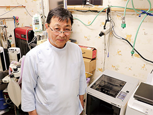
Dr. Seiichi Takemura, Director of Komoro Animal Hospital
Conclusive fact in introducing 3DFF-222
- Mimaki Support and Maintenance
- Good Price Point
From One Book to the 3D World
"I pursue my interests to the fullest extent" -Dr. Takemura
I came up with the idea of outputting CT data on a 3D printer when I found a book called “Medical Image 3D Modeling and 3D Printer Utilization Guide” by Maki Sugimoto, a physician and entrepreneur. I realized that by transforming the data taken by CT into three dimensions and outputting it with a 3D printer, bones and organs can be checked three-dimensionally, and I thought that this should be used for animal treatment as well.
After purchasing a 3D printer online, I tried various things on my own, but it didn't work out. It is easy to buy online, but there is no support for the equipment and the instruction manual is written in English, so it was often difficult to know what caused the output. At that time, I read an article in a local newspaper that Mimaki released a small 3D printer, the 3DFF-222.
I knew of Mimaki, but until then I thought it was a company that only manufactures large inkjet printers. I knew Mimaki could provide me with support, and since they started selling small 3D printers, I immediately inquired online.
Materialization of Desires Through Thorough Support
When the sales person first came, they brought a lot of samples output by the 3DFF-222. They wanted to understand my needs and if they could be met with this printer. It was easy to purchase in terms of price, and it was compact, with no space issues. After that, they responded promptly to various inquiries and have been really helpful.
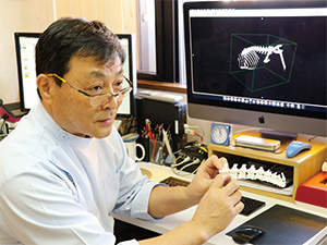
For example, I wanted to check inside the bone of a herniated disc, a common problem with dogs, but when I output the CT image as is with the 3DFF-222, the inside of the bone was closed and I was not able to see what I needed to. So I tried to divide the bone in half with the data and output it so that I could see inside, but it didn't work. When I contacted Mimaki with this issue, I was told that it would be possible to output with a free 3D CAD. As a result, the shape, orientation, and size of the protruding bone can be checked thoroughly from the half-length bone model, as well as how to insert a scalpel and sharpen the bone. I am now able to have a good simulation before surgery.
The biggest advantage of simulations with 3D models is that the burden on animals is reduced. Preoperative simulation can reduce the operation time and minimize the number of incisions and eliminate unnecessary incisions. When explaining to the owner, I can talk with something concrete and visible, which reduces their uncertainty as well.
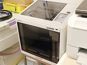
3DFF-222
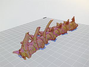
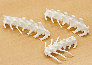
Animal skeleton 3D model created soon after purchasing the 3DFF-222
3DFF-222 Streamlines Inspection to Surgery
The output time of 3DFF-222 is about 4 to 6 hours for dog and cat bones. It's now possible to send the CT data to the 3DFF-222 in the evening to be finished by morning, we can chose an accurate treatment option and examination, explain it to the owner, and perform the operation very efficiently.
Some service providers will send you 3D models if you send CT data, but this is a huge time loss. The great advantage of having your own printer is that everything can be done at your own pace.
In addition, explanations using 3D models have enabled the owner to understand the importance of CT examinations during medical treatment. In the case of animals, CT and surgery cost a great deal. By using CT and 3D models together, we can understand the precise treatment method, and show us that preoperative simulations will enable the minimization of invasive surgery, thereby deepening our understanding of CT examinations.
It has been about half a year after the introduction of our printer. Although there are still only few cases where 3DFF-222 has been utilized, based on the data that we have accumulated so far, we intend to further utilize 3DFF-222 for endoscopic treatment.
Skeletal model for preoperative simulation
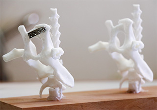
Confirming the position of fixing bolts to fractured bones
The Potential of Veterinary Medicine with 3D Printers
3D models are very useful for treatment, but it would be better if you could actually cut it with a high-speed drill for simulation. The current material melts with heat, so I can't be certain of the feeling on my hands when cutting the bones or how it will cut. If there would be a material whose hardness, texture, etc. are close to those of bone, more realistic simulations could be possible.
In addition, if 3D models could be made with a material that could be safely implanted into a living body, it would be possible to create a customized structure for each individual, such as the shape and curve of each bone, minimalizing the invasiveness and creating safer treatment.
For example, when a dog or cat fractures its pelvis, reduction surgery is performed to fix the pelvic bone with a metal plate and bolts. I must do it on the spot. This increases the surgery time, and once the bone is fixed, you will have to do another surgery to remove the bolts and metal plates. Two operations are a significant burden for small dogs and cats. If we could do a single operation with 3D structures, it would be fastened with the best curved plates and bolts, and once it's absorbed, it wouldn't need to be removed. It may be difficult, but I think veterinary medicine will have great developments if it becomes possible.
Mimaki's 3D printer is not for medical use, but I hope they will continue to work on product development as a bridge between the industrial and veterinary fields.
Komoro Animal Hospital
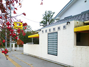
Komoro Animal Hospital is located in Nagano prefecture, Japan.
Opened in 1983, they moved to their current location in 1998. For 36 years, it has been treating pets such as dogs, cats, rabbits and small birds.
In 2006, its sister hospital “Usuda Pet Clinic” was opened. Utilizing 3D technology, which has already been put into practical use in such medical fields as dental care, it enables minimally invasive surgery by using a combination of CT scans and 3D printers.
Facilities for radiography, electrocardiography, blood pressure measurement, skin camera inspection, etc.

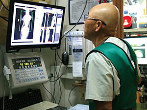
Surgery with CT image diagnosis
<Introduced products>
FFF system Desktop 3D Printer : 3DFF-222
User profile
- NameKomoro Animal Hospital
- IndustryVeterinarian
- Address2728-1 Mikageshinden, Komoro-shi, Nagano, Japan
- Phone number+81-267-23-6644
- URLhttps://www.komoro-animal.jp/
Related articles

- 3DUJ-553 reduces the time and effort required for 3D modeling & effective use of full-color output greatly expands applications : The University of Tokyo

- How Mimaki’s Full Color 3D Printer is Revolutionizing the Footwear Design Process

- Bringing Adobe Substance 3D Models to Life Using a Mimaki Full Color 3D Printer! - The printed models were given to the talented creators at CREAPOLE, a design school in France -